


- News
25 August 2015
Kwansei Gakuin University uses Renishaw Raman microscope to study defects in SiC
Professor Noboru Ohtani and colleagues in the Department of Nanotechnology for Sustainable Energy of the School of Science and Technology at Japan's Kwansei Gakuin University have reported on using Renishaw's inVia confocal Raman microscope for micro-Raman imaging to help show the influence of heavily doped nitrogen donors on the defect formation in silicon carbide (T Takahashi et al, 'Structural and electrical characterization of the initial stage of physical vapour transport growth of 4H-SiC crystals', Materials Science Forum vols 821-823 (2015) p90).
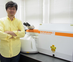 Picture: Kwansei Gakuin University's professor Noboru Ohtani, with his Renishaw inVia Raman microscope.
Crystallographic defects in 4H-SiC epitaxial wafers (such as dislocations and stacking faults) limit the commercialization of SiC devices and must therefore be eliminated or reduced to levels lower than some critical density. The department's research goal is to establish SiC crystal growth processes that can produce large, ultra-high-quality SiC epitaxial wafers. To achieve this, they try to clarify the cause and formation mechanism of crystallographic defects in SiC bulk crystal and epitaxial film.
Crystallographic defects give rise to residual stresses in the crystals. The stresses can occur through a variety of mechanisms in 4H-SiC crystals. For example, temperature gradients in the grown crystals, which are a primary driving force for crystal growth, lead to plastic deformation of the crystals during the growth and/or cooling process. This deformation results in residual stresses when the crystals are cooled to room temperature. The spatial variation of stresses in the crystals can be measured using Renishaw's inVia confocal Raman microscope. These measurements provide information about the formation of defects during physical vapour transport (PVT) growth and chemical vapour deposition (CVD) processes. This information can be used to improve the crystal growth process.
Ohtani's laboratory also uses high-resolution x-ray diffraction (HR-XRD) to characterize stress distribution. Raman microscopy provides complementary information to the HR-XRD data but with much higher spatial resolution. "The key benefit is the ultra-high speed data acquisition system, which results in a higher sensitivity to measuring stresses in the materials compared to other Raman systems," commented Ohtani regarding the inVia system.
www.scientific.net/MSF.821-823.90